Guide to Thalassiosira minima

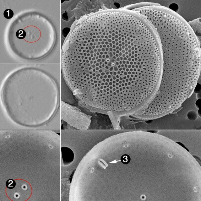
- Cells small
- Two central fultoportulae
- Rimoportula oriented radially
Cells are small. Although electron microscopy is required for a definitive determination of the species, the number and position of both the
marginal and subcentral fultoportulae and the orientation of the rimoportula can often be revealed with careful light microscopy. Two central, closely-spaced fultoportulae are present. The internal opening of the marginal rimoportula is elongated in a radial orientation and can be distinguished using LM.
 Diatoms of North America